Health Tests | 15 min read
Electrocardiograph (EKG): Type, Result and Process
Medically reviewed by
Table of Content
Key Takeaways
- ECG stands for electrocardiogram and is a painless, non-invasive tes
- An ECG test helps detect irregular heart rate patterns and rhythms
- An ECG scan can indicate hypertension or a previous heart attack
A simple test to check your heart's rhythmic and electrical activity is an ECG test.
ECG stands for electrocardiogram. It is the go-to test for detecting heart ailments. An ECG scan is a safe and non-invasive procedure. Doctors recommend this for people facing chest pain, breathing difficulties, high blood pressure, dizziness, palpitations, or any irregularities in the heartbeat.
If you have diabetes, high cholesterol, smoke, or have a family history of heart illness, an ECG test is recommended.
Apart from providing information about heart rate and rhythm, an ECG also detects if the heart is enlarged due to hypertension or if you have had any recent heart attacks. An ECG test is effective in detecting the following conditions.
- Cardiomyopathy due to the thickening of the heart walls
- Heart attacks due to the blockage of blood supply to the heart
- Arrhythmias due to irregular heartbeat
- Coronary heart disease is due to the presence of fatty substances blocking the blood supply to the heart.
Here is an overview of the ECG scan process and its different types and readings.
Types Of ECG Tests
In addition to a standard electrocardiogram (ECG), your doctor may suggest the following:
Holter Monitor:
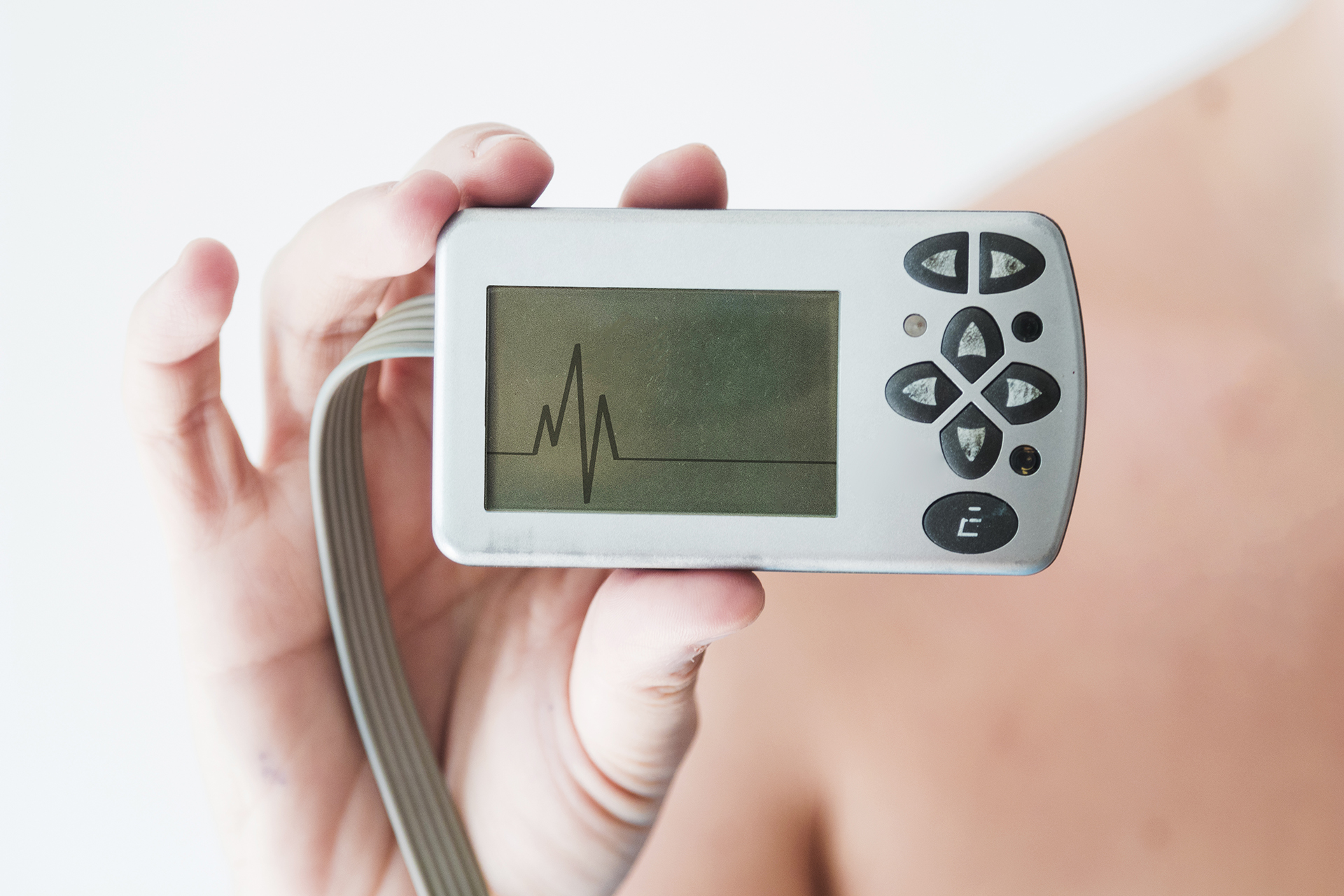
It is a small, portable device that records the heart's electrical activity over a period of time, typically 24 to 48 hours. It is often used if you experience symptoms such as heart palpitations, an irregular heartbeat, or reduced blood flow to the heart. During the test, electrodes will be attached to your chest, arms, and legs, and you will be able to go about your normal activities while wearing the monitor.
Event Monitor:
This portable device is used to record the heart's electrical activity over a longer period, typically weeks or even months. It is often used when you experience symptoms occasionally or irregularly. With an event monitor, you will need to push a button or activate the device whenever you experience symptoms, and it will record the electrical activity of your heart at that time. Some event monitors are designed to automatically record whenever they detect an abnormal rhythm in your heart.
Interpreting The Results Of ECG
After undergoing an electrocardiogram (ECG), your doctor will review and discuss the test results with you. If the results are normal, you may not need to undergo any additional testing. However, if the results indicate an abnormal ECG, your doctor may recommend further testing or another ECG to determine the cause of your symptoms. To determine the appropriate treatment for your symptoms, your doctor will review the information recorded during your electrocardiogram (ECG) and check for any potential heart problems, which include:
Heart rhythm:
One of the things they will check for is heart rhythm. With an ECG, your doctor can detect arrhythmias or irregular heart rhythms. An arrhythmia can occur when any part of the heart's electrical system fails to function properly. Some medications, including amphetamines, beta-blockers, over-the-counter allergy and cold medications, and cocaine, can also trigger arrhythmias.
Heart rate:
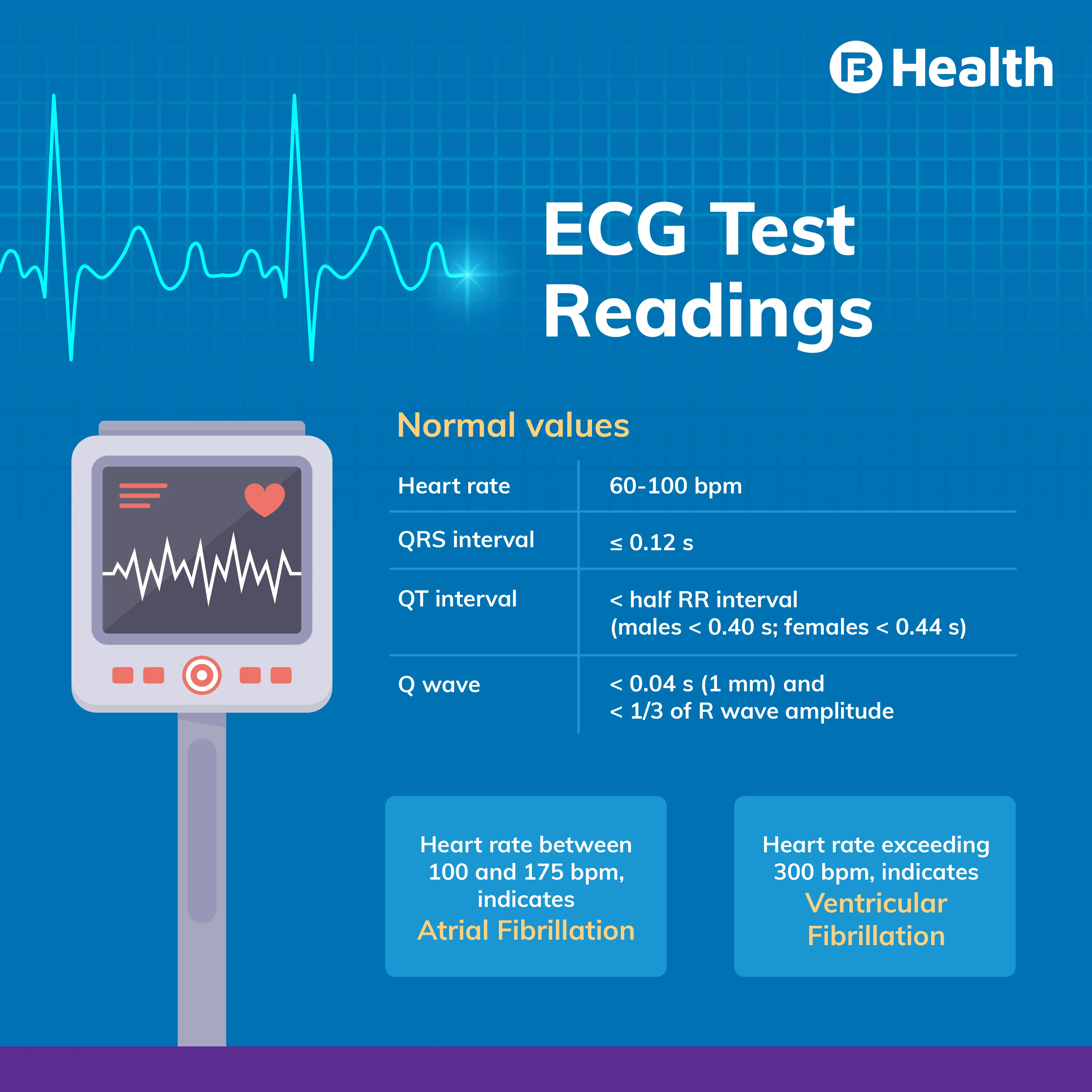
It is typically measured by checking the pulse and can be diagnosed using an electrocardiogram (ECG) when the pulse is also irregular or fast to measure accurately. With an ECG, your doctor can diagnose tachycardia, a condition in which the heart beats faster than normal (usually more than 100 times per minute), or bradycardia, a condition in which the heart beats slower than normal (usually less than 60 times per minute). An ECG can help determine the cause and appropriate treatment for an abnormal heart rate.
Heart Attack:
An electrocardiogram (ECG) can be used to detect a heart attack, whether it has already occurred or is imminent, by analysing the ECG patterns. These patterns can reveal the location of damaged heart tissue and the extent of the damage caused by the heart attack. An ECG is useful for diagnosing heart attacks and determining the appropriate treatment.
Structural Abnormalities of Heart:
An electrocardiogram (ECG) can be used to detect structural abnormalities of the heart, such as the enlargement of heart chambers or walls, as well as various heart diseases. By analysing the ECG patterns, doctors can identify potential problems with the heart's structure and determine the appropriate course of treatment. A range of factors, including genetics, lifestyle, and underlying medical conditions, can cause structural abnormalities in the heart. An ECG is essential for identifying these abnormalities and determining the best course of action to address them.
Insufficient Oxygen and Blood Supply to The Heart:
During an electrocardiogram (ECG) test, if you are experiencing symptoms, your doctor can determine whether inadequate blood flow to the heart is causing your symptoms, such as chest pain. For example, unstable angina, a type of chest pain resulting from reduced blood flow to the heart, can be diagnosed using an ECG. If your doctor detects any problems on your ECG, they may recommend further testing to determine the cause and whether treatment is necessary. A range of factors can cause insufficient oxygen and blood supply to the heart, including blocked or narrowed blood vessels, heart valve problems, and heart muscle damage. An ECG can help identify these issues and guide treatment decisions.
Types of ECG Devices
An electrocardiogram (EKG) is a test that tracks the heart's electrical activity for a short period of time. This means that it may not be able to detect heart irregularities that only occur infrequently, similar to how internet issues may not be apparent when a technician is trying to diagnose the problem.
One way to diagnose intermittent heart problems is by using a Holter monitor. This device is worn for 24 to 48 hours and continuously records the heart's electrical activity during that time. Another option is an event monitor, which can be worn for a week or longer. With this device, the wearer may need to press a button to start recording whenever they experience symptoms. Both monitors can provide valuable information to your healthcare provider to identify and address any potential issues with your heart.
How To Read An EKG
The human heart has its own internal pacemaker called the sinoatrial node, which generates electrical signals to initiate the heartbeat. These signals can be recorded and monitored using an electrocardiogram (EKG or ECG). An EKG machine reads the heart's electrical activity as it contracts and relaxes with each heartbeat, providing valuable information about the heart's health.
During an EKG, the healthcare provider will evaluate the strength and duration of the electrical signals and the time intervals between different peaks and waves representing the electrical impulses. The first wave, known as the "P wave," is produced by the heart's upper chambers (atria), where the heartbeat originates. The second wave, known as the QRS complex, is generated by the lower chambers of the heart (ventricles). The third wave, called the "T wave," represents the heart's resting or recovery phase after a beat.
What is an ECG vs. EKG?
An electrocardiogram (EKG or ECG) is a test that evaluates the electrical activity of the heart. It is used to detect any abnormalities or issues with the heart's rhythm and function. The term EKG is derived from the German word "Elektrokardiogramm," which uses the letter "k" in place of "c" in both parts of the word. It is important to note that an EKG is different from an echocardiogram, which is a type of ultrasound that produces images of the heart as it beats. An echocardiogram utilises high-frequency sound waves to create a detailed picture of the heart and its structures, while an EKG uses electrodes to measure the heart's electrical activity. Both tests can provide important information about the health of the heart, but they serve different purposes and use different technologies.
When Would An EKG Be Used?
An electrocardiogram (EKG or ECG) is a useful tool that allows healthcare providers to assess the heart's electrical activity and detect any potential issues or abnormalities. There are several reasons why a healthcare provider may order an EKG, including:
- Evaluating the heart rhythm to determine if it is normal or if the patient has an arrhythmia (abnormal heart rhythm)
- Diagnosing poor blood flow to the heart muscle (ischemia) due to coronary artery disease
- Detecting a heart attack
- Identifying abnormalities of the heart, such as heart chamber enlargement and abnormal electrical conduction
- Diagnosing heart damage or heart failure
- Determining whether the patient is fit for an upcoming surgery
In addition to these purposes, an EKG can also be used to monitor the heart's condition in patients who have received a pacemaker, started taking medication for heart disease, or experienced a heart attack. By regularly tracking the heart's electrical activity, healthcare providers can ensure that the heart is functioning properly and make any necessary adjustments to treatment plans.
Symptoms You Can Diagnose With An EKG
Many different symptoms may prompt a healthcare provider to order an electrocardiogram (EKG or ECG) for a patient. An EKG test measures the heart's electrical activity and allows healthcare providers to assess the heart's rhythm and function. Some common reasons for performing an EKG include the following:
Chest pain:
If a patient experiences chest pain, an EKG can help the healthcare provider determine whether a heart issue, such as a heart attack or angina, is causing the pain
Shortness of breath:
Difficulty breathing can be a sign of a heart problem, and an EKG can help the healthcare provider identify the cause
Tiredness: Persistent fatigue or tiredness may be a sign of an underlying heart condition, and an EKG can help the healthcare provider diagnose the problem
Dizziness:
Dizziness or lightheadedness can be a symptom of an abnormal heart rhythm, and an EKG can help the healthcare provider determine if this is the case
A flutter or skip in a heartbeat:
If a patient experiences a fluttering or skipping sensation in their heartbeat, an EKG can help the healthcare provider diagnose the cause
A fast heartbeat:
A rapid heart rate (tachycardia) can be a sign of a problem in the heart, and an EKG can help the healthcare provider determine the cause and recommend the appropriate treatment
Overall, an EKG is useful for diagnosing and treating various heart-related conditions. It is often performed in response to certain symptoms that may indicate a problem with the heart.
Who Performs an EKG?
An electrocardiogram (EKG or ECG) is a diagnostic test used to monitor the heart's electrical activity. It is often ordered or performed by a cardiologist, a healthcare provider specialising in diagnosing and treating heart conditions. However, other healthcare providers may also order or perform an EKG, especially in emergency situations, such as when a patient is being treated in an ambulance or the emergency room. An EKG can be administered in various settings, including a healthcare provider's office, a hospital, or an outpatient facility.
How do I Prepare for an EKG Test?
An electrocardiogram (EKG or ECG) is a non-invasive test. It is typically performed in a healthcare provider's office, a hospital, or an outpatient facility. Before taking an EKG, you can eat and normally drink without making special preparations. However, there are a few things to consider for getting dressed on the day of the test:
- Avoid using oily or greasy skin creams and lotions. These substances can interfere with the electrodes making good contact with your skin, which is necessary for accurate test results
- Avoid wearing full-length hosiery. Electrodes need to be placed directly on the legs, so it is essential to wear clothing that allows easy access to your skin
- Wear a shirt that you can easily remove. The electrodes for the EKG will need to be placed on your chest, so wearing a shirt that can be easily removed or opened is helpful
Overall, it is important to follow these guidelines to ensure that the EKG can be performed smoothly and accurately. If you have any questions about preparing for an EKG, it is always a good idea to speak with your doctor.
What Type Of Results Do You Get, And What Do The Results Mean?
Some of the things that an EKG may reveal include the following:
An irregular, fast, or slow heart rhythm:
An EKG can detect any deviations from a normal heart rhythm and help the healthcare provider diagnose and treat conditions such as arrhythmias
A past or present heart attack:
An EKG can detect changes in the heart's electrical activity that may be indicative of a heart attack, even if the attack occurred in the past
Cardiomyopathy or an aneurysm:
An EKG can identify changes in the structure of the heart, such as thickened heart walls (cardiomyopathy) or stretched-out areas (aneurysms)
Poor blood flow to the heart:
An EKG can reveal problems with blood flow to the heart, such as ischemia due to coronary artery disease
Heart failure:
An EKG can detect signs of heart failure, such as abnormal electrical activity or changes in the heart's structure
What to Expect During an EKG
An electrocardiogram (EKG or ECG) is a quick, painless, and harmless diagnostic test that measures the heart's electrical activity. During an EKG, you can expect the following steps:
- You will be asked to change into a gown
- Using gel, a technician will attach about ten soft electrodes to your chest, arms, and legs. These electrodes are connected to wires that are attached to the EKG machine
- If the areas where the electrodes are attached are not shaved, the technician may shave them to ensure good contact
- You will be asked to lie still on the table and breathe normally. It is important to refrain from talking during the test
- The machine will record your heart's electrical activity and display the results on a graph
- Once the test is complete, the electrodes will be removed and discarded
- The entire procedure typically takes about 10 minutes
An EKG is a simple test that provides valuable information about the heart's health. It is quick, painless, and harmless and can be an important tool in diagnosing and treating heart-related conditions.
Interpreting the Results of an EKG
If your electrocardiogram (EKG or ECG) test results are normal, your healthcare provider will typically review the results with you during your appointment or at a follow-up visit. In this case, you can expect to receive a full explanation of the results and any recommendations for further care or monitoring.
On the other hand, if the EKG results appear abnormal or suggest the presence of any health problems, your healthcare provider should contact you as soon as possible to discuss the findings and options for improving your heart's condition. It is essential to follow your healthcare provider's recommendations to ensure the best possible outcomes for your health.
Some signs of abnormalities that may show up in an EKG include the following:
Irregular heartbeat:
An EKG can detect deviations from a normal heart rhythm, such as arrhythmias
Heart defects: An EKG can identify heart defects such as an enlarged heart, a lack of blood flow, or congenital disabilities
Electrolyte problems:
An EKG can detect problems with electrolytes, which are substances that help regulate the body's fluid balance and maintain normal heart function
Chamber dilation or hypertrophy:
An EKG can identify changes in the size or shape of the heart's chambers, such as dilation (enlargement) or hypertrophy (thickening)
Abnormal electrical conduction:
An EKG can detect abnormalities in the way electricity is moving through the heart
Blocked arteries or coronary artery disease:
An EKG can reveal problems with blood flow to the heart, such as blockages in the arteries (coronary artery disease)
If the EKG test shows signs of any abnormalities, your healthcare provider will discuss options to improve your heart's condition. This may include prescribing medication or suggesting lifestyle changes such as modifying your diet or increasing your physical activity.
ECG Test Process
Normally, you don’t need to fast before undergoing an ECG. However, it is advisable to provide vital information to your doctor regarding your medications or if you have a pacemaker implanted in your chest.
Electrodes or small sticky sensors are put on your arms, chest, and legs before the test. These sensors connect to a recording machine that detects electrical currents the heart generates.
ECG Tests are of three main types to check your heart rate activity:
- A resting ECG
- Stress or exercise ECG
- Ambulatory ECG
- You have to lie down for a resting ECG. The stress test or exercise ECG records your heart rate when you walk on a treadmill or ride an exercise bike. Ambulatory ECG records your heart rate when connected to a small machine on your waist. Your heart rate is monitored for 24 hours at home. You can continue with your daily routine with this home option.
implies that while your readings may not be precisely normal, they are not significantly abnormal either. An abnormal ECG indicates a defect or abnormality in your heart.
Symptoms and Causes of Ventricular Fibrillation
Ventricular fibrillation is a type of arrhythmia that occurs due to abnormal heart rhythm. As a result of abnormal heart signals, lower heart chambers or ventricles tremble unnecessarily. This stops the heart from pumping blood to the rest of the body. Ventricular fibrillation requires immediate medical attention as it can lead to sudden cardiac arrest.
Some of the signs you need to watch out for includes the following:
- Dizziness
- Nausea
- Rapid beating of the heart
- Chest pain
A ventricular fibrillation ECG is done along with chest X-ray, echocardiogram, CT scan, MRI scan, and blood tests to diagnose this condition. An ECG helps reveal if there are irregularities in your heartbeat. Usually, a rate of 300 to 400 bpm indicates ventricular fibrillation. [3]

Causes And Risk Factors Of Atrial Fibrillation
Atrial fibrillation is yet another heart ailment that can affect your heartbeat rate. However, in this condition, the irregular beating causes the heart's upper chambers or atria to beat so fast that they begin to quiver. Consequently, your ventricles also begin to beat irregularly.
Atrial fibrillation is also quite dangerous as it increases the risk of heart failure and stroke. Both atria and ventricles work in unison so that heart pumps blood properly. However, in this condition, the heartbeat rate increases somewhere around 100 to 175 bpm.
Few symptoms of this condition include the following:
- Faster heartbeats
- Chest pain
- Uneven pulse
- A rapid fluttering of the heart
The ideal way to check this condition is to undergo an atrial fibrillation ECG. This way, your doctor can detect if there is a problem with the rhythm of your heartbeat.
While an ECG helps you assess the risk factors associated with the heart, it is essential to maintain an active and healthy lifestyle to protect yourself from heart ailments. Try to follow a well-balanced diet and strive to keep your weight under control. Keep an eye on heart problems' risk factors and symptoms and ensure you get an ECG test when needed. Book all lab tests with ease on Bajaj Finserv Health so you can nip heart problems in the bud.
References
- https://www.nhs.uk/conditions/electrocardiogram/
- https://www.betterhealth.vic.gov.au/health/conditionsandtreatments/ecg-test
- https://www.ncbi.nlm.nih.gov/books/NBK537120/
- https://www.premierheartandveincare.com/can-detect-blocked-arteries-ecg/
- https://www.webmd.com/heart-disease/atrial-fibrillation/ss/slideshow-af-overview
- https://www.mayoclinic.org/diseases-conditions/ventricular-fibrillation/diagnosis-treatment/syc-20364524
- https://www.mayoclinic.org/diseases-conditions/ventricular-fibrillation/symptoms-causes/syc-20364523
- https://www.mayoclinic.org/tests-procedures/ekg/about/pac-20384983
- https://choosingwiselycanada.org/ecg-electrocardiogram/
- https://www.webmd.com/heart-disease/electrocardiogram-ekgs
- https://www.hopkinsmedicine.org/health/treatment-tests-and-therapies/electrocardiogram
Disclaimer
Please note that this article is solely meant for informational purposes and Bajaj Finserv Health Limited (“BFHL”) does not shoulder any responsibility of the views/advice/information expressed/given by the writer/reviewer/originator. This article should not be considered as a substitute for any medical advice, diagnosis or treatment. Always consult with your trusted physician/qualified healthcare professional to evaluate your medical condition. The above article has been reviewed by a qualified doctor and BFHL is not responsible for any damages for any information or services provided by any third party.

